Meningioma
Meningioma is a primary brain tumour originating in the meninges, the three-layered membrane enclosing the brain and spinal cord. Most cases are WHO Grade I (benign) and grow slowly over years. Approximately 37% of all diagnosed primary brain tumours are meningiomas, making them the most common intracranial tumour type.
Understanding Meningioma
Although the majority of meningiomas are non-cancerous, their growth inside the rigid skull inevitably compresses surrounding brain structures. Over time, a slowly expanding tumour can press against the cerebral cortex, cranial nerves, or venous sinuses, producing neurological symptoms that worsen as the tumour enlarges. The challenge with meningioma is that by the time a patient notices symptoms, the tumour may have been present and growing for a decade or more.
Early specialist evaluation transforms the outlook: small tumours caught before they cause permanent damage offer far more treatment options, shorter recovery times, and higher success rates. For patients diagnosed overseas, Istanbul provides access to world-class meningioma expertise with waiting times measured in days rather than months.
Treatment Options for Meningioma
View All ProceduresBrain Aneurysm Treatment in Turkey
A brain aneurysm (cerebral aneurysm) is a weakened, balloon-like bulge in the wall of a blood vessel in the brain. Most aneurysms are small, cause no symptoms, and are discovered incidentally during imaging for other conditions. However, if an aneurysm ruptures, it causes a subarachnoid haemorrhage (bleeding around the brain), a life-threatening emergency with a mortality rate of approximately 30–50%.
arrow_forwardBrain Tumour Surgery in Turkey
Brain tumour treatment demands two things above all: an experienced neurosurgeon and access to the full range of treatment modalities. Istanbul offers both, Vellum Select curates two neurosurgeons whose combined experience exceeds 30,000 treated patients and whose complementary expertise spans open microsurgery, Gamma Knife radiosurgery, neuronavigation, awake craniotomy, and intraoperative imaging.
arrow_forwardGamma Knife Radiosurgery in Turkey
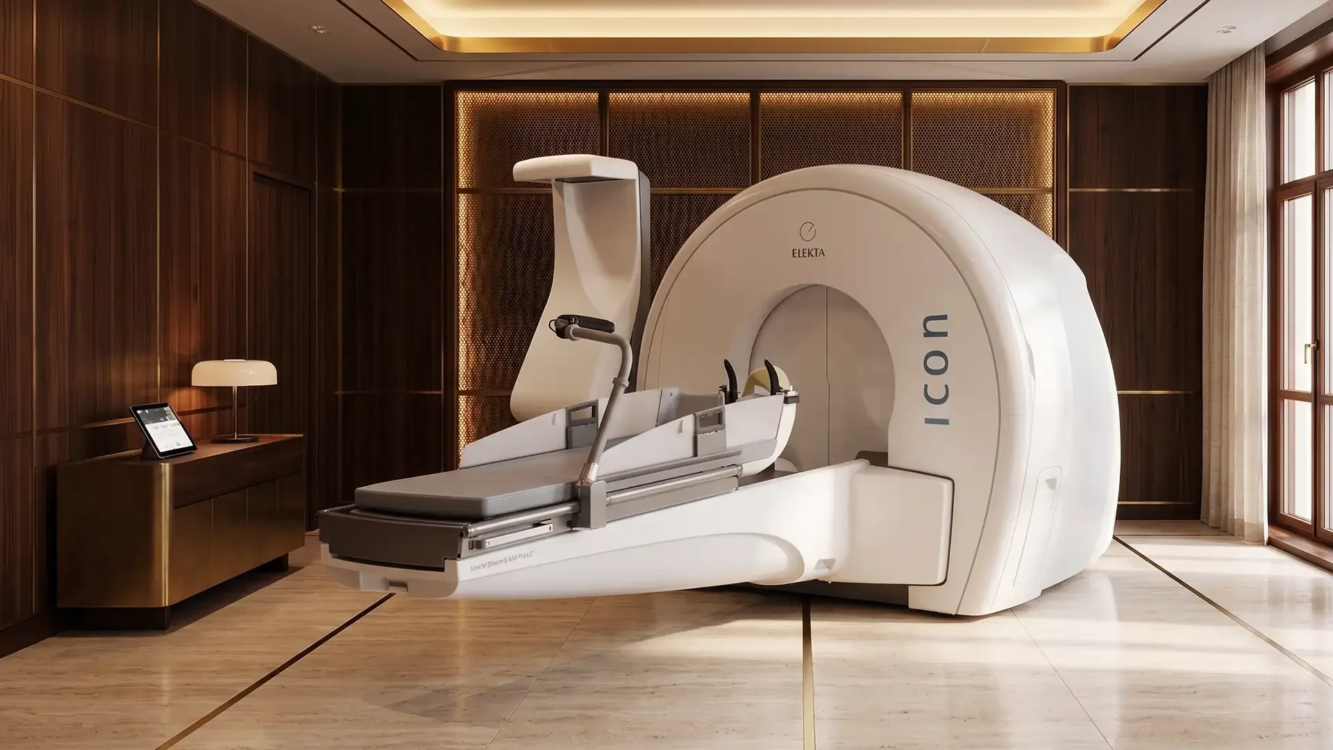
Gamma Knife radiosurgery is a non-invasive stereotactic procedure that delivers 192 intersecting cobalt-60 radiation beams to a single focal point inside the brain with sub-millimetre accuracy. Each individual beam is too weak to damage the tissue it passes through; only at the convergence point does the accumulated dose become therapeutically powerful enough to destroy a tumour or lesion, with no incision, no cutting, and no general anaesthetic required.
arrow_forwardSymptoms of Meningioma
Meningioma symptoms vary widely depending on the tumour's size and location within the skull. The most common symptoms include persistent or worsening headaches (often worse in the morning), seizures in patients with no prior history of epilepsy, vision changes including blurring or loss of peripheral vision, weakness or numbness in the arms or legs, memory loss or difficulty concentrating, changes in personality or mood, and hearing loss or tinnitus for tumours near the acoustic nerve.
Many small meningiomas are discovered incidentally during imaging performed for unrelated reasons and cause no symptoms at all. These cases are often managed with active surveillance rather than immediate intervention.
Diagnostic Pathways
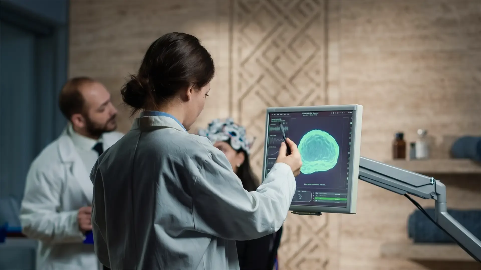
MRI with contrast enhancement is the definitive imaging modality for meningioma. These tumours have a characteristic appearance on contrast-enhanced MRI, with a homogeneously enhancing mass and a "dural tail" sign. Most meningiomas can be diagnosed by imaging alone without a biopsy.
CT scanning is used to evaluate calcification within the tumour, which is common in meningiomas, and to assess bone involvement or hyperostosis of the overlying skull.
Angiography or MRA may be used to assess the tumour's blood supply, particularly for large or skull base meningiomas where preoperative embolisation may reduce surgical bleeding.
Pathological grading is performed when tissue is obtained through biopsy or surgical resection. The WHO classification grades meningiomas from Grade I (benign) to Grade III (malignant), which determines the subsequent treatment strategy and surveillance protocol.
Advanced Treatment Options at Vellum Select
The optimal treatment for meningioma depends on tumour size, location, WHO grade, patient symptoms, and overall health. Vellum Select offers the full spectrum of meningioma management through Prof. Dr. Türker Kılıç, one of the world's most experienced meningioma surgeons.
Gamma Knife Radiosurgery
For small to medium-sized meningiomas (typically under 3 cm) that are either asymptomatic or causing mild symptoms, Gamma Knife Radiosurgery is often the preferred approach. Gamma Knife uses 192 focused radiation beams to arrest tumour growth with sub-millimetre accuracy, requiring no incision and no hospital admission. Tumour control rates for Grade I meningiomas exceed 95% at 5 years. The procedure is performed as a single outpatient session, and most international patients return home within 48 hours.
Craniotomy and Microsurgical Resection
For larger meningiomas causing significant mass effect or neurological symptoms, or for tumours accessible enough for complete removal, Brain Tumour Surgery (craniotomy) remains the gold standard. Prof. Dr. Kılıç's microsurgical technique aims for complete resection (Simpson Grade I or II), which offers the lowest recurrence rates. Post-operative Gamma Knife is often used to treat any residual tumour at the skull base, combining the strengths of both approaches.
Active Surveillance
Small, asymptomatic meningiomas in elderly patients or those with significant surgical risk may be managed with serial MRI monitoring. Prof. Dr. Kılıç defines individualised surveillance intervals based on tumour growth rate and patient factors, ensuring intervention is recommended only when the benefit clearly outweighs the risk.
To discuss your meningioma diagnosis with Prof. Dr. Türker Kılıç, view his profile or contact Vellum Select to arrange a consultation.
Related Articles
Gamma Knife vs. Open Brain Surgery: A Clinical Comparison
Yes, Gamma Knife radiosurgery is a highly effective, non-invasive alternative to open brain surgery for specific deep-seated brain tumors, AVMs, and neuralgias. While open surgery is required for large tumors causing immediate intracranial pressure, Gamma Knife offers sub-millimeter precision without incisions, general anesthesia, or prolonged ICU stays.

The Real Cost of Medical Treatment in Turkey: A Price Comparison Guide (2026)
How much does medical treatment in Turkey actually cost? This comprehensive price guide compares dental veneers, implants, rhinoplasty, and neurosurgery costs in Turkey vs. the UK and Europe, explaining the structural reasons behind the savings.

Is Turkey Safe for Medical Tourism? An Honest Guide for European Patients
European patients consistently ask the same question before booking: is Turkey actually safe? This guide answers directly, with facts about medical accreditation, city safety, patient rights, and how to identify a clinic you can trust.