Arteriovenous Malformation
Arteriovenous malformation (AVM) is a congenital vascular anomaly in which arteries connect directly to veins without an intervening capillary bed, creating a high-flow nidus that risks spontaneous haemorrhage. AVMs are the leading cause of intracerebral haemorrhage in young adults, with an annual rupture risk of 2–4 per cent.
Understanding Brain AVMs
In a normally developed brain, arteries deliver oxygenated blood through progressively smaller vessels — arterioles and capillaries — before reaching the venous system. In an AVM, this capillary network is absent; high-pressure arterial blood flows directly into thin-walled veins that are not designed to withstand such pressure. Over time, the abnormal vessels dilate, the nidus may enlarge, and the risk of rupture increases. Each haemorrhage carries a 10–15 per cent mortality rate and a 30–50 per cent risk of permanent neurological deficit.
Even unruptured AVMs produce symptoms through a mechanism called vascular steal, where the high-flow shunt diverts blood away from surrounding healthy brain tissue, causing progressive neurological deficits and seizures. Treatment aims to completely obliterate the nidus, eliminating both the rupture risk and the steal phenomenon. Istanbul offers access to expert multidisciplinary AVM management, combining Gamma Knife radiosurgery with advanced endovascular techniques at a fraction of Western costs.
Treatment Options for Arteriovenous Malformation
View All ProceduresSymptoms of Brain AVM
Approximately 50 per cent of AVMs first present with intracranial haemorrhage, producing sudden severe headache, nausea, vomiting, and focal neurological deficits corresponding to the haemorrhage location. Seizures are the presenting symptom in 20–25 per cent of patients, particularly with superficial frontal or temporal AVMs. Chronic headaches, progressive neurological deficits from vascular steal, and pulsatile tinnitus are common in unruptured AVMs. An audible bruit over the skull or eye may be detected on auscultation in high-flow lesions.
Diagnostic Pathways
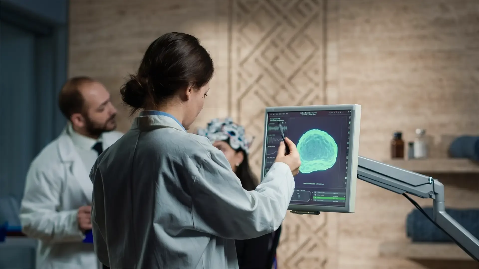
CT angiography (CTA) is the first-line imaging for suspected acute haemorrhage and can identify the AVM nidus and its arterial supply. MRI/MRA provides detailed anatomical localisation of the nidus and its relationship to eloquent brain regions. Digital subtraction angiography (DSA) remains the gold standard for AVM grading — the Spetzler-Martin classification (Size × Eloquence × Venous Drainage) determines the treatment strategy and surgical risk. DSA provides dynamic visualisation of arterial feeders, the nidus, and draining veins essential for treatment planning.
Advanced Treatment Options at Vellum Select
Vellum Select provides comprehensive AVM management through Prof. Dr. Türker Kılıç, integrating Gamma Knife radiosurgery, microsurgical resection, and endovascular techniques.
Gamma Knife Radiosurgery
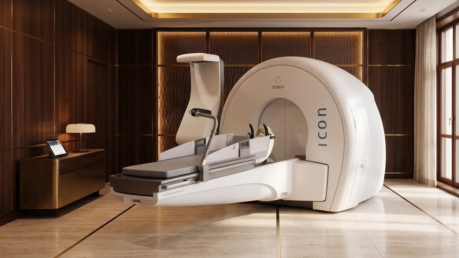
For small to medium-sized AVMs (under 3 cm) located in deep or eloquent brain regions, Gamma Knife Radiosurgery in Turkey is the preferred modality. The treatment delivers precisely focused radiation to the nidus, inducing progressive endothelial proliferation and vessel wall thickening that leads to complete obliteration over 2–3 years. Obliteration rates reach 80 per cent at three years for AVMs under 3 cm. During the latency period, the rupture risk is not eliminated but is reduced, and patients undergo serial MRI surveillance. The procedure is outpatient, non-invasive, and international patients typically return home within 48 hours.
Microsurgical Resection
For superficial AVMs in non-eloquent areas — Spetzler-Martin grades I and II — microsurgical resection offers immediate cure. Combined approaches using pre-operative embolisation followed by Gamma Knife are employed for complex, large, or high-grade AVMs where single-modality treatment is insufficient.
To discuss your AVM diagnosis with Prof. Dr. Türker Kılıç, view his profile or contact Vellum Select to arrange a consultation.
Related Articles
Gamma Knife vs. Open Brain Surgery: A Clinical Comparison
Yes, Gamma Knife radiosurgery is a highly effective, non-invasive alternative to open brain surgery for specific deep-seated brain tumors, AVMs, and neuralgias. While open surgery is required for large tumors causing immediate intracranial pressure, Gamma Knife offers sub-millimeter precision without incisions, general anesthesia, or prolonged ICU stays.

The Real Cost of Medical Treatment in Turkey: A Price Comparison Guide (2026)
How much does medical treatment in Turkey actually cost? This comprehensive price guide compares dental veneers, implants, rhinoplasty, and neurosurgery costs in Turkey vs. the UK and Europe, explaining the structural reasons behind the savings.

Is Turkey Safe for Medical Tourism? An Honest Guide for European Patients
European patients consistently ask the same question before booking: is Turkey actually safe? This guide answers directly, with facts about medical accreditation, city safety, patient rights, and how to identify a clinic you can trust.