Craniotomy (Brain Surgery) in Turkey
Open Brain Surgery: Precision Neurosurgery in Istanbul
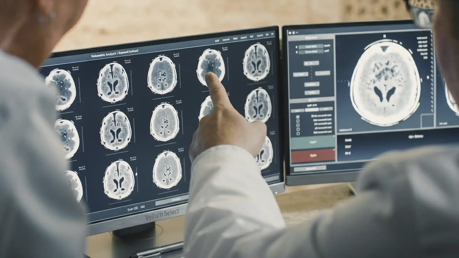
Craniotomy is the foundation of modern neurosurgery. When a brain tumour, aneurysm, or vascular malformation is too large, too deep, or too threatening for radiosurgery alone, opening the skull remains the standard of care—and often the only curative option. The difference between an uneventful recovery and a complication is the experience of the surgeon interpreting real-time anatomy through a surgical microscope.
Prof. Dr. Türker Kılıç has performed over 30,000 cranial procedures across his career, bridging microsurgery and Gamma Knife radiosurgery to offer each patient the optimal treatment pathway. International patients travel to Istanbul for access to his Harvard-trained expertise at costs 60 to 70 per cent below UK and German private neurosurgery rates.
Conditions This Treatment Addresses
Frequently Asked Questions
About Craniotomy (Brain Surgery) in Turkey
What is a craniotomy?
A craniotomy is the surgical opening of the skull to access the brain. A section of bone (bone flap) is temporarily removed, the intracranial procedure is performed under an operating microscope, and the bone flap is secured back in place with titanium plates and screws at closure.
How long does recovery take after a craniotomy?
Hospital stay is typically 3 to 7 days. Most patients return to sedentary work within 4 to 6 weeks and full physical activity within 8 to 12 weeks. Recovery time varies depending on the size and location of the lesion and the patient's preoperative condition.
How much does a craniotomy cost in Turkey?
Craniotomy in Istanbul costs approximately £8,000–£18,000 depending on procedure complexity, compared to £25,000–£50,000 in the UK. The price includes preoperative imaging, the surgery, ICU stay, and hospitalisation at a JCI-accredited facility.
What are the risks of a craniotomy?
Risks include infection (1–2%), bleeding, swelling, neurological deficit (varies by lesion location), seizure, and CSF leak. Prof. Kılıç uses intraoperative neuromonitoring to map and preserve eloquent brain areas and has a complication rate significantly below international benchmarks due to his volume of over 30,000 procedures.
Will I need to shave my head?
Only a narrow strip of hair along the planned incision line is shaved. The rest of your hair remains untouched. In many cases, the incision is planned within a natural skin crease and will be hidden once hair regrows. Prof. Kılıç's closures are designed to minimise visible scarring.
Can I fly home after a craniotomy?
Most international patients are cleared to fly 7 to 10 days after surgery, once sutures are removed and the wound is healing well. Prof. Kılıç's team reviews a post-operative MRI to confirm no complications before authorising air travel.
What Is a Craniotomy?
A craniotomy begins with the patient under general anaesthesia. The scalp is incised and reflected, and a series of burr holes are drilled through the skull. A surgical saw connects these holes, freeing a bone flap that is lifted away to expose the dura mater. The dura is opened, and the brain is accessed. Under the operating microscope (providing 6–40× magnification with stereoscopic depth perception), the surgeon performs the intracranial procedure: tumour resection, aneurysm clipping, or AVM excision.
At closure, the dura is sutured watertight, the bone flap is secured with titanium plates and screws, and the scalp is closed. The bone flap typically heals and fuses over 6 to 12 months.
Conditions Treated by Craniotomy
Brain Tumours
Craniotomy is the primary treatment for gliomas (including glioblastoma), meningiomas, acoustic neuromas, brain metastases, pituitary adenomas, and other intracranial tumours. The goal is maximal safe resection—balancing complete tumour removal against preservation of neurological function. Intraoperative neuromonitoring and awake craniotomy techniques map eloquent brain areas during surgery.
Read more about Brain Tumour Surgery in Turkey →
Brain Aneurysms
A cerebral aneurysm is a weakened, ballooning segment of an artery wall at risk of rupture and catastrophic subarachnoid haemorrhage. Microsurgical clipping—placing a titanium clip across the aneurysm neck to exclude it from the circulation—performed via craniotomy remains the most durable treatment with the lowest long-term rebleed rate.
Arteriovenous Malformations (AVMs)
For large or ruptured AVMs where Gamma Knife is insufficient or too slow, microsurgical resection via craniotomy provides immediate, definitive cure. The surgeon isolates and divides feeding arteries, dissects the nidus, and divides draining veins in a precise sequence that avoids intraoperative haemorrhage.
Craniotomy Cost in Turkey vs. the UK and Europe
| Procedure | UK Cost | Turkey (Istanbul) Cost |
|---|---|---|
| Brain Tumour Craniotomy | £25,000–£45,000 | £8,000–£15,000 |
| Aneurysm Clipping | £30,000–£50,000 | £10,000–£18,000 |
| AVM Resection | £25,000–£45,000 | £8,000–£16,000 |
Prices include preoperative imaging (MRI, angiography where required), the surgical procedure with intraoperative neuromonitoring, ICU stay, and inpatient hospitalisation. Prof. Kılıç operates at BAU Medical Park Göztepe Hospital, a JCI-accredited facility.
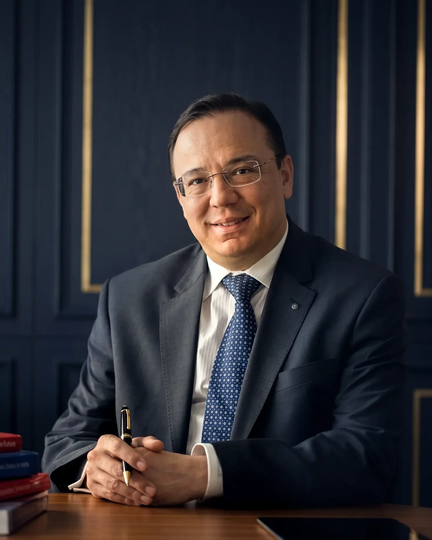
Your Surgeon: Prof. Dr. Türker Kılıç
Vellum Select's curated craniotomy specialist is Prof. Dr. Türker Kılıç, who trained at Hacettepe University and Harvard University and has performed over 30,000 cranial procedures. He holds an H-index of 40 and has published more than 200 scientific papers. Prof. Kılıç is the only scientist in Turkey elected to both the European Academy of Sciences and Arts and the World Academy of Art and Science.
His dual expertise in microsurgical craniotomy and Gamma Knife radiosurgery means treatment recommendations are never constrained by the limitations of a single technique. He will recommend craniotomy when it offers superior cure potential and Gamma Knife when it provides equivalent results with less risk.
View Prof. Dr. Kılıç's full profile →
Craniotomy vs. Gamma Knife Radiosurgery
| Factor | Craniotomy | Gamma Knife |
|---|---|---|
| Incision | Scalp incision + bone flap | None |
| Anaesthesia | General | Local (frame pins only) |
| Hospital Stay | 3–7 days | Same day or 1 night |
| Recovery | 4–8 weeks | 1–2 days |
| Result | Immediate tumour removal | Gradual tumour control (months–years) |
| Best For | Large or mass-effecting lesions | Small-medium lesions, deep locations |
These are complementary techniques, not competitors. Prof. Kılıç's practice encompasses both, ensuring each patient receives the approach best suited to their specific pathology.
Journey to Recovery
Day 1: Craniotomy and ICU Admission
The surgery is performed under general anaesthesia and typically lasts 3 to 6 hours depending on the complexity of the lesion. After closure, you are transferred directly to the neurosurgical intensive care unit (ICU) for continuous neurological monitoring. A head bandage is in place and a drain may be present. Family members are updated by Prof. Kılıç after the procedure.
Days 2 to 3: ICU to Ward Transfer
Once stable and alert, you are transferred from ICU to a private neurosurgical ward room. The head drain (if placed) is removed. Pain is managed with intravenous then oral analgesia. You will begin sitting up and taking short walks with assistance by day 3. A post-operative MRI confirms the extent of resection.
Days 4 to 7: Hospital Recovery and Discharge Planning
Mobility improves daily. Sutures or staples remain in place. The neurosurgical team reviews your discharge plan, including wound care instructions, activity restrictions, and medication. Most international patients are discharged to their hotel by day 5 to 7 with a companion and a follow-up appointment for suture removal.
Days 7 to 10: Suture Removal and Initial Follow-Up
Return to the clinic for suture or staple removal and a wound check. Prof. Kılıç reviews the post-operative MRI and pathology report (if tumour). At this point, most international patients are cleared to fly home, provided they are neurologically stable and have no signs of infection or CSF leak.
Weeks 2 to 6: Early Recovery at Home
Avoid heavy lifting, strenuous activity, and activities that increase intracranial pressure (straining, bending). Headache and fatigue are normal and gradually subside. You may gradually resume work if it is sedentary. A follow-up MRI is typically scheduled at 6 to 12 weeks, which can be arranged locally and shared with Prof. Kılıç's team.
Months 3 to 6: Full Recovery
By 3 months, most patients have returned to their normal routine. The bone flap is healing and the scalp incision is barely visible within the hairline. Ongoing surveillance imaging (MRI every 6–12 months) monitors for recurrence. Prof. Kılıç's team remains available for remote consultation.
Learn more about Craniotomy (Brain Surgery) in Turkey
Your journey to better health starts here. Fill in the form and a dedicated consultant will contact you shortly.
Global Concierge
+90 555 709 02 07