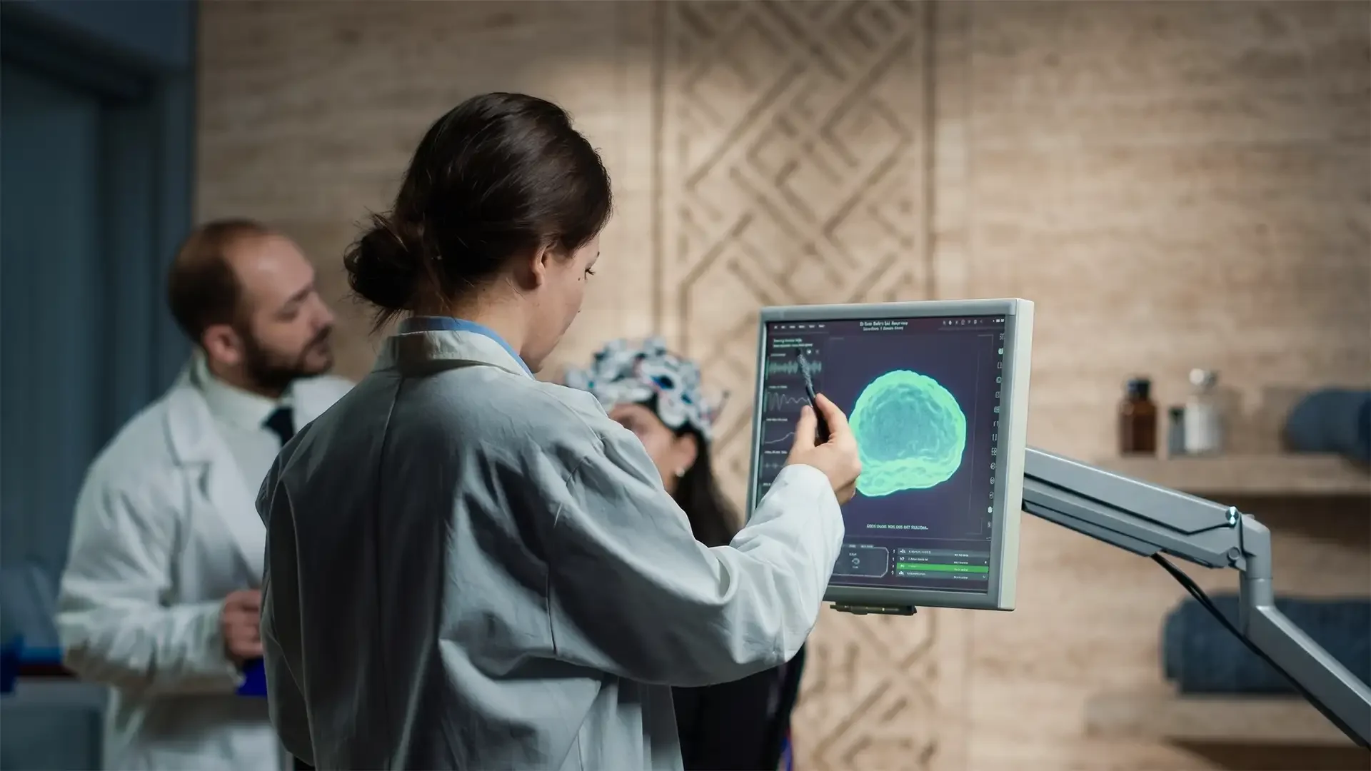
Brain Aneurysm
A brain aneurysm is a pathological focal dilation of a cerebral artery wall, most commonly at bifurcation points in the Circle of Willis. The wall structure is compromised by loss of the tunica media and internal elastic lamina, creating a thin-walled sac that may rupture, causing subarachnoid haemorrhage — a medical emergency with 50 per cent mortality.
Understanding Brain Aneurysms
The majority of cerebral aneurysms — approximately 3 per cent of the population carries one — remain asymptomatic and undetected throughout life. The danger lies in the 0.5–1 per cent annual rupture risk, which increases with aneurysm size, smoking, hypertension, and family history. Ruptured aneurysms produce subarachnoid haemorrhage (SAH), a catastrophic event with an immediate mortality of 10–15 per cent and a further 25 per cent mortality within the first 30 days. Survivors face significant morbidity from vasospasm, rebleeding, and hydrocephalus.
Treatment of unruptured aneurysms — either surgical clipping or endovascular coiling — eliminates the rupture risk and provides peace of mind. For patients diagnosed overseas, Istanbul offers access to elite neurovascular surgeons and internationally accredited hospitals with waiting times measured in days, at 50–60 per cent below US and UK private healthcare costs.
Treatment Options for Brain Aneurysm
View All ProceduresTratamiento del Aneurisma Cerebral en Turquía
Un aneurisma cerebral es una dilatación anormal de la pared de una arteria en el cerebro. La mayoría son asintomáticos, pero la rotura causa una hemorragia subaracnoidea potencialmente mortal.
arrow_forwardCraneotomía (Cirugía Cerebral) en Turquía
Una craneotomía es la apertura quirúrgica del cráneo para acceder al cerebro y extirpar tumores, clipar aneurismas, resecar malformaciones arteriovenosas (MAV) o descomprimir tejido cerebral edematoso. Se extrae temporalmente un colgajo óseo, se realiza el procedimiento intracraneal bajo microscopio y se fija el hueso nuevamente al cerrar.
arrow_forwardSymptoms of Brain Aneurysm
Most unruptured brain aneurysms cause no symptoms and are discovered incidentally during imaging for unrelated reasons. Larger aneurysms may produce mass effect symptoms: a posterior communicating artery aneurysm can cause a third nerve palsy with ptosis and diplopia; a giant aneurysm can present with headache, visual disturbance, or seizure. A sentinel headache — sudden and severe but self-limiting — may precede a major rupture by days to weeks and is frequently misdiagnosed as migraine. Rupture presents as the classic "thunderclap" headache: the worst headache of the patient's life, of instantaneous onset, often accompanied by nausea, vomiting, neck stiffness, photophobia, and loss of consciousness.
Diagnostic Pathways
CT head without contrast is the first-line investigation for suspected SAH — hyperdense blood in the basal cisterns is visible within hours of rupture. CT angiography (CTA) provides detailed characterisation of the aneurysm morphology — size, neck width, dome-to-neck ratio, and relationship to parent and perforating vessels. Digital subtraction angiography (DSA) remains the gold standard for treatment planning, particularly for complex or wide-necked aneurysms. Lumbar puncture is performed when CT is negative but clinical suspicion remains high — xanthochromia of the cerebrospinal fluid is diagnostic of recent SAH.
Advanced Treatment Options at Vellum Select
Vellum Select offers comprehensive aneurysm management through Prof. Dr. Türker Kılıç, a leading neurovascular surgeon with extensive experience in both microsurgical and endovascular techniques.
Microsurgical Clipping
For aneurysms with favourable anatomy — particularly middle cerebral artery bifurcation aneurysms and those in young patients — Craniotomy (Brain Surgery) in Turkey with microsurgical clip placement provides definitive and permanent exclusion of the aneurysm. The procedure involves a craniotomy, microscopic dissection of the aneurysm neck, and placement of a titanium clip that permanently seals the aneurysm from the circulation. Cure rates exceed 98 per cent for unruptured aneurysms.
Endovascular Treatment
For aneurysms amenable to a less invasive approach, Brain Aneurysm Treatment in Turkey offers coiling, stent-assisted coiling, or flow diversion (such as the Pipeline embolisation device) via a catheter advanced from the femoral artery. No incision, no craniotomy, and recovery is typically 24–48 hours in hospital. Flow diversion has revolutionised the treatment of large and giant aneurysms previously considered inoperable.
To discuss your brain aneurysm diagnosis with Prof. Dr. Türker Kılıç, view his profile or contact Vellum Select to arrange a consultation.